On the 50th anniversary of American chemist Pal Laterbur’s groundbreaking discovery of magnetic resonance imaging (MRI), scientists have achieved a major milestone in medical imaging. Researchers from Duke University’s Center for In Vivo Microscopy, along with scientists from the University of Tennessee Health Science Center, University of Pennsylvania, University of Pittsburgh and Indiana University, have developed an MRI that is 64 million times sharper than current technology.
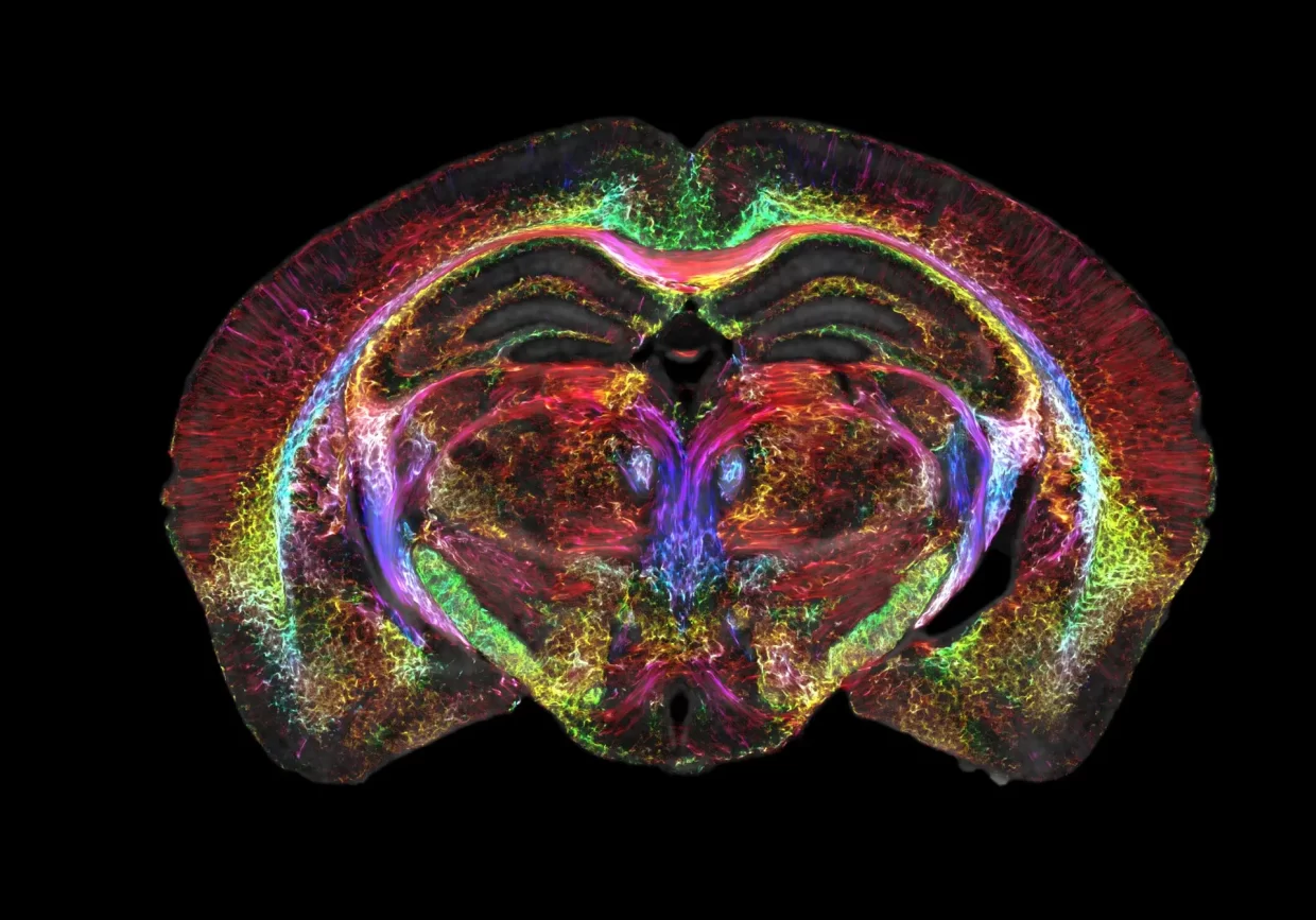
This MRI is capable of capturing images with a resolution of 5 microns, or five-thousandth of a millimeter, per voxel – the 3D version of a pixel. This level of detail allows for a much more detailed view of the brain than current MRI technology, which is only able to spot tumors. The new MRI can display organization and connectivity in the brain, as demonstrated in the accompanying video. This technology could revolutionize medical imaging and provide unprecedented insight into the inner workings of the brain.
This breakthrough in MRI imaging allowed researchers to capture a single mouse brain in unprecedented detail. This level of imaging could provide a greater understanding of how brains change with age, diet, and neurodegenerative diseases such as Alzheimer’s.
Lead author G. Allan Johnson, professor of radiology, physics, and biomedical engineering at Duke, said, “It is something that is truly enabling. We can start looking at neurodegenerative diseases in an entirely different way.”
The team used a powerful 9.4-Tesla magnet, 100 times stronger than the magnets used in standard scans, along with a set of gradient coils and a supercomputer equivalent to 800 laptops. This combination of cutting-edge technology made the achievement possible, after almost 40 years of work at the Center for In Vivo Microscopy.
Shedding Light on Neurodegenerative Disease Progression
Using advanced imaging techniques, researchers have been able to gain a greater understanding of how neurodegenerative diseases progress over time. By combining MRI visuals with light sheet microscopy, they were able to label specific groups of cells and observe how the brain-wide connectivity of mice changed with age and genetic makeup. The images also revealed the breakdown of neural networks caused by Alzheimer’s disease.
This research opens the door to further technological development that could capture the human brain in such detail, allowing us to better understand how tissue changes with age and what interventions could be used to prevent degeneration.
According to Dr. Johnson of the National Institute of Aging, modest dietary and drug interventions can lead to animals living 25% longer, so the question is whether their brains remain intact during this extended lifespan. With the capacity to look at this in greater detail, we can translate the findings directly into the human condition.
The groundbreaking study has been published in the prestigious peer-reviewed scientific journal, Proceedings of the National Academy of Sciences (PNAS).